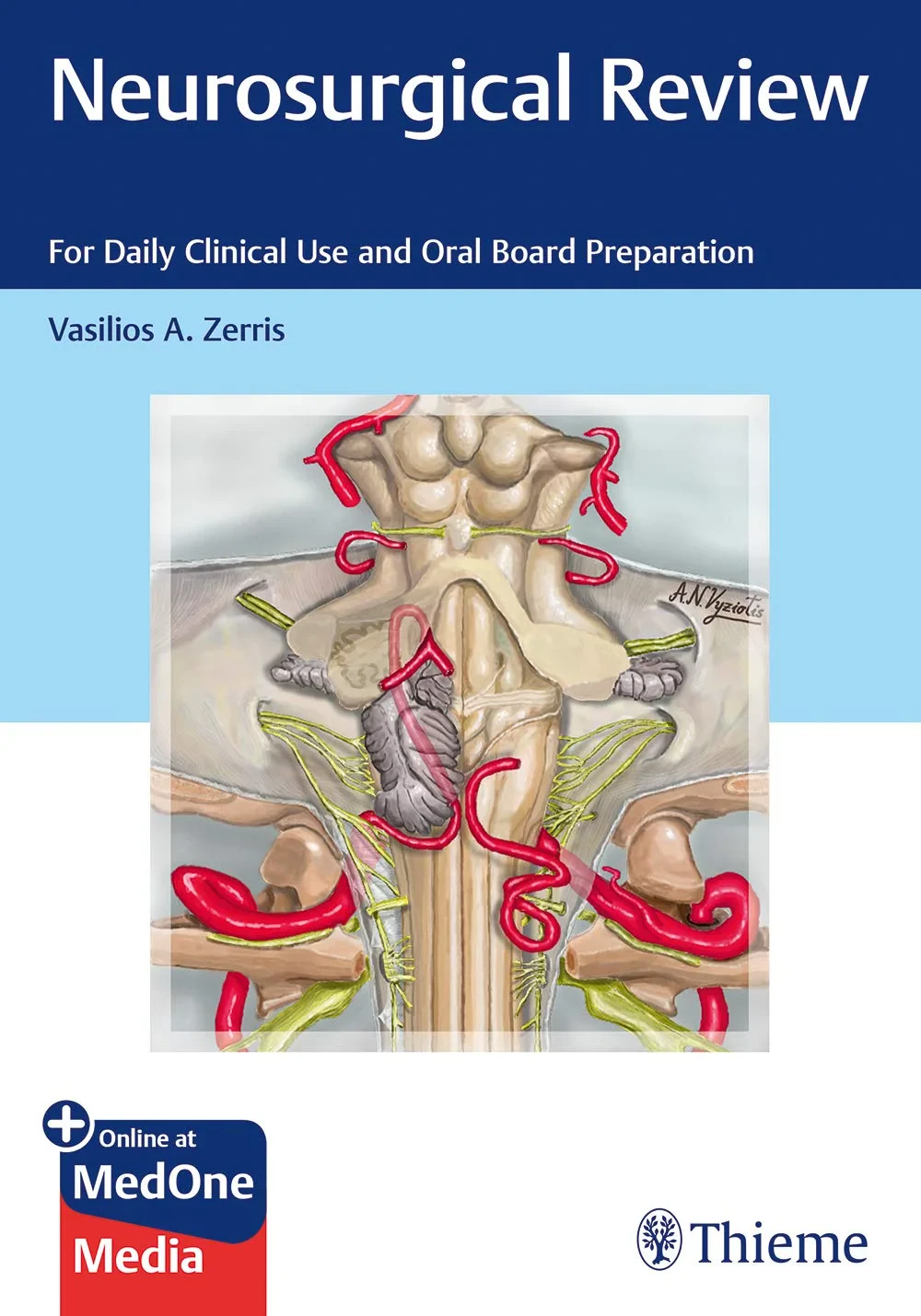
Description:
Robust ABNS exam prep and didactic review of the entire spectrum of neurosurgery from A to Z
The American Board of Neurological Surgery oral examination has undergone periodic review and revision over the years, with a new format instituted in spring
2017. This review book is specifically geared to the new format. The ABNS oral examination process is relevant, rigorous, and of value to the neurosurgical specialty and the public, ensuring neurosurgeons meet the highest standards of practice.
Neurosurgical Review: For Daily Clinical Use and Oral Board Preparation by Vasilios A. Zerris and distinguished contributors is a multimodal and a visually rich prep tool for the ABNS exam. The resource provides a unique approach to studying and melding online didactic materials with audio-enhanced charts. Readers can use the material as a complete online exam prep course with audio, or use the print version as a quick reference guide.
Key Features:
Charts and schematics provide an excellent learning tool and study prep
The high yield and easy to memorize format helps readers "visualize" knowledge
Audio files enhance the ability to create a mental framework, thereby increasing comprehension and retention of content
Cases presented at the end of each chapter focus primarily on core material tested in the general neurosurgery ABNS exam session taken by all candidates irrespective of their declared subspecialty
This is an essential textbook for neurosurgical residents, fellows, and practitioners prepping for the ABNS boards. It also serves as a user-friendly refresher of fundamental knowledge all neurosurgeons need to know.
Table of Contents:
1 Peripheral Nerves
1. 1 Diagnostic Approach for Peripheral Nerve Lesions (Table
1. 1)
1. 2 Neuropathies
1.
2. 1 Nerve pathologies depending on number and location of nerves involved (Table
1. 2a)
1.
2. 2 Other classifications for polyneuropathies (Table
1. 2b)
1.
2. 3 Causes for neuropathies (Table
1. 2c)
1.
2. 4 Peripheral neuropathy versus radiculopathy (Table
1. 2d)
1. 3 Peripheral Nerve Injuries
1.
3. 1 Basics (Table
1. 3a)
1.
3. 2 Regeneration: (Table
1. 3b)
1.
3. 3 Mechs of nerve injury (Table
1. 3c)
1.
3. 4 Grading of Nerve Injuries (Table
1. 3d)
1.
3. 5 Peripheral Nerve Injury Grading Systems (Table
1. 3e)
1.
3. 6 Management of Peripheral Nerve Injury (Table
1. 3f)
1.
3. 7 Timing of Nerve Exploration and Repair Rule of 3s (Table
1. 3g)
1.
3. 8 Algorithm of Timing of Nerve Surgery (Table
1. 3h)
1.
3. 9 Peripheral Nerve Repair: Surgical Pearls (Table
1. 3i)
1. 4 Brachial Plexus
1.
4. 1 Brachial Plexus Injuries (Table
1. 4a)
1.
4. 2 Brachial Plexus Injuries: Trunks (Table
1. 4b)
1.
4. 3 Brachial Plexus Injuries: Cords (Table
1. 4c)
1.
4. 4 Dx of Preganglionic Injury (Nerve Root Avulsion) (Table
1. 4d)
1.
4. 5 Surgical Approach Based on Defining Clinical Level of Lesion (Table
1. 4e)
1.
4. 6 Supraclavicular Approach (Table
1. 4f)
1.
4. 7 Infraclavicular Approach (Table
1. 4g)
1.
4. 8 Neurogenic Thoracic Outlet Syndrome (Table
1. 4h)
1.
4. 9 Parsonage–Turner Syndrome (Table
1. 4i)
1. 5 Entrapment Neuropathies
1.
5. 1 Median Nerve [(C5), C6, C7, C8, T1] Sites of Compression (Table
1. 5a)
1.
5. 2 Median Nerve Entrapment Sites: Motor and Sensory Deficits (Table
1. 5b)
1.
5. 3 Clinical Syndromes and Findings: Median Nerve Injury/Compression in Arm (Table
1. 5c)
1.
5. 4 Management for Injury of Median Nerve in Arm (A, B) (Table
1. 5d)
1.
5. 5 Key surgical steps for median nerve exposure in arm (A, B) (Table
1. 5e)
1.
5. 6 Clinical Syndromes and Findings: Median Nerve Entrapment in FA (Table
1. 5f)
1.
5. 7 Clinical Syndromes and Findings: Median Nerve / AIN Injury/Compression in FA (Table
1. 5g)
1.
5. 8 Management for Entrapment of Median Nerve/AIN at Distal Arm/Elbow/Proximal FA (C, D) (Table
1. 5h)
1.
5. 9 Key Surgical Steps for Decompression of Median n./AIN at Distal Arm/Elbow/Proximal FA (C, D) (Table
1. 5i)
1.
5. 10 Clinical Syndromes and Findings: Median Nerve Entrapment in Carpal Tunnel (Table
1. 5j)
1.
5. 11 Management for Entrapment of Median Nerve at Carpal Tunnel (E) (Table
1. 5k)
1.
5. 12 Key Surgical Steps for Median Nerve Decompression at the Carpal Tunnel (E) (Table
1. 5l)
1. 6 Ulnar Nerve
1.
6. 1 Ulnar nerve (C7, C8, T1) (Table
1. 6a)
1.
6. 2 Ulnar nerve entrapment sites: motor and sensory deficits (Table
1. 6b)
1.
6. 3 Clinical Syndromes and Findings: Ulnar Nerve Injury/Compression in Arm (Table
1. 6c)
1.
6. 4 Management for Injury of Ulnar Nerve in Arm (A)(Table
1. 6d)
1.
6. 5 Key Surgical Steps for Ulnar Nerve Exposure in Arm (A) (Table
1. 6e)
1.
6. 6 Clinical Syndromes and Findings: Ulnar Nerve Injury/Compression in Elbow (Table
1. 6f)
1.
6. 7 Clinical Syndromes and Findings: Ulnar Nerve Injury/Compression in FA (Table
1. 6g)
1.
6. 8 Management for Injury of Ulnar Nerve Injury/Compression in Elbow/FA (B, C, D) (Table
1. 6h)
1.
6. 9 Key Surgical Steps for Ulnar Nerve Decompression in Elbow/FA (B, C, D) (Table
1. 6i)
1.
6. 10 Clinical Syndromes and Findings: Ulnar Nerve Injury/Compression at Guyon’s Tunnel (Table
1. 6j)
1.
6. 11 Management for Ulnar Nerve Compression/Injury at Guyon’s Tunnel (Table
1. 6k)
1.
6. 12 Key Surgical Steps for Ulnar Nerve Decompression in Guyon’s Tunnel (Table
1. 6l)
1. 7 Radial Nerve
1.
7. 1 Radial nerve (C5, C6, C7, C8, [T1]) (Table
1. 7a)
1.
7. 2 Radial nerve entrapment sites: motor and sensory deficits(Table
1. 7b)
1.
7. 3 Clinical Syndromes and Findings: Radial Nerve Compression/Injury in Arm (Table
1. 7c)
1.
7. 4 Management for Radial Nerve Injury in Arm
1.
7. 5 Clinical Syndromes and Findings: Radial Nerve Compression/Injury at Antecubital Fossa/FA (Table
1. 7f)
1.
7. 6 Clinical Syndromes and Findings: Radial Nerve Compression/Injury at FA (Table
1. 7g)
1.
7. 7 Management for Entrapment of Radial Nerve/PIN at Elbow/FA (Table
1. 7h)
1.
7. 8 Approaches for radial nerve decompression in radial tunnel syndrome and PIN decompression (C, D) (Table
1. 7i)
1. 8 Sciatic Nerve (L4, L5, S1, S2, S3) and Tibial Nerve (L4, L5, S1, S2, S3) (Table
1. 8a)
1.
8. 1 Common Peroneal Nerve (L4, L5, S1, S2) (Table
1. 8b)
1.
8. 2 Sciatic Nerve (Table
1. 8c)
1.
8. 3 Sciatic Nerve Entrapment Sites: Motor and Sensory Deficits (Table
1. 8d-f)
1.
8. 4 Clinical Syndromes and Findings: Sciatic Nerve Compression/Injury at Buttock (Table
1. 8g)
1.
8. 5 Clinical Syndromes and Findings: Sciatic Nerve Compression/Injury at Thigh (Table
1. 8h)
1.
8. 6 Management for Compression/Injury of Sciatic Nerve at Buttock/Thigh (A, B) (Table
1. 8i)
1.
8. 7 Key Surgical Steps for Sciatic Nerve Exposure at Buttock (A) (Table
1. 8j)
1.
8. 8 Key Surgical Steps for Sciatic Nerve Exposure at Thigh (B) (Table
1. 8k)
1.
8. 9 Clinical Syndromes and Findings: Common Peroneal n. and Tibial n. Compression/Injury at Knee/Leg (Table
1. 8l)
1.
8. 10 DDx of Foot Drop (Table
1. 8m)
1.
8. 11 Management for Compression/Injury of CPN at Knee/Leg (D) (Table
1. 8n)
1.
8. 12 Key Surgical Steps for CPN Exposure at Knee/Leg (Table
1. 8o)
1.
8. 13 Clinical Syndromes and Findings: Compression of Tibial and CPN at Foot (Table
1. 8p)
1.
8. 14 Management for Compression/Injury of Tibial Nerve and its Branches (Table
1. 8q)
1.
8. 15 Key Surgical Steps for Exposure of Tibial Nerve and its Branches at Knee/Leg/Foot
1. 9 Meralgia Paresthetica (Table
1. 9)
1. 10 Postures (Table
1. 10)
1. 11 Peripheral Nerve Tumors
1.
11. 1 Classification of Peripheral Nerve Tumors (Table
1. 11a)
1.
11. 2 Peripheral Nerve Tumors (Table
1. 11b)
1.
11. 3 Schwannoma Versus Neurofibroma (Table
1. 11c)
1.
11. 4 Management (Table
1. 11d)
1.
11. 5 Malignant Peripheral Nerve Sheath Tumor (MPNST) (Table
1. 11e)
1. 12 Abbreviations
1. 13 Cases
1.
13. 1 Brachial Plexus Stab Wound
1.
13. 2 Ulnar Neuropathy
1.
13. 3 Carpal Tunnel Syndrome
2 Spine and Spinal Cord
2. 1 Key Myotomes and Dermatomes and Their Main Function (Table
2. 1)
2. 2 Spine Injuries
2.
2. 1 Management of Cervical Spine Fractures (Table
2. 2a)
2.
2. 2 Management of Thoracic and Lumbar Spine Fractures (Table
2. 3)
2.
2. 3 Management of Sacral Spine Fractures (Table
2. 4)
2.
2. 4 Penetrating Spine Injuries (Table
2. 5)
2. 3 Management of Degenerative Cervical Spine Disease
2. 4 Other Spine or Spinal Cord Conditions (Table
2. 7)
2. 5 Spinal Infections (Table
2. 8)
2. 6 Management of Degenerative Thoracic and Lumbar Spine Disease (Table
2. 9)
2. 7 Spinal Instrumentation Placement (Table
2. 10)
2.
7. 1 Ten Spinal Instrumentation Rescue and Salvage Techniques (Table
2. 11)
2. 8 Pediatric Spine and Spinal Cord Injury Guidelines (Table
2. 12)
2. 9 Scoliosis (Table
2. 13)
2. 10 Tumors of Spine and Spinal Cord (Table
2. 14)
2. 11 Cases
2.
11. 1 Cervical Spine Fracture with Perched Facet
2.
11. 2 Lumbar Disc Herniation with Cauda Equina Syndrome
2.
11. 3 Cervical Spondylotic M yelopathy—Central Cord Syndrome
2.
11. 4 Spinal Epidural Abscess
3 Vascular Neurosurgery
3. 1 Aneurysms
3.
1. 1 Nonruptured Aneurysms (Table
3. 1a)
3.
1. 2 Ruptured Aneurysms: Spontaneous Subarachnoid Hemorrhage (Table
3. 1d)
3.
1. 3 Vascular Spasm
3.
1. 4 Clipping Versus Coiling
3.
1. 5 Aneurysm Surgery: General Recommendations
3.
1. 6 Aneurysm Surgery: Details per Location
3.
1. 7 Miscellaneous
3. 2 Intracranial Hemorrhage
3.
2. 1 Spontaneous Intracerebral Hemorrhage (American Heart Association/American Stroke Association Guidelines 2015) (Table
3. 2a)
3.
2. 2 STICH trial (The International Surgical Trial in Intracerebral Hemorrhage) (Table
3. 2b)
3.
2. 3 STICH II (Table
3. 2c)
3. 3 Carotid Disease
3.
3. 1 Studies
3.
3. 2 Treatment Options Stenting (Table
3. 3d)
3. 4 Stroke
3.
4. 1 Stroke rate (Table
3. 4a)
3.
4. 2 Stroke syndromes (Table
3. 4b)
3.
4. 3 Vertebral insufficiency (5Ds) (Table
3. 4c)
3.
4. 4 Ischemic stroke (American Heart Association/American Stroke Association guidelines 2013 and revision 2015, Adapted) (Table
3. 4d)
3. 5 Moyamoya Disease
3.
5. 1 Moyamoya disease (Table
3. 5a)
3.
5. 2 Moyamoya Disease: Suzuki Stages in DSA (Table
3. 5b)
3.
5. 3 Management of Moyamoya Disease (Table
3. 5c)
3. 6 Brain Arteriovenous Malformations
3.
6. 1 Spetzler–Martin scale (Table
3. 6a)
3.
6. 2 AVM rupture rates (Table
3. 6b)
3.
6. 3 Multiple AVMs (Table
3. 6c)
3.
6. 4 ARUBA trial (A Randomized Trial of Unruptured Brain Arteriovenous malformations) (Table
3. 6d)
3.
6. 5 Treatment based on Spetzler–Martin scale (Table
3. 6e)
3.
6. 6 Timing for other treatment modalities after AVM embolization (Table
3. 6f)
3.
6. 7 Surgery for AVMs
3. 7 Spinal Arteriovenous Malformations
3.
7. 1 Artery of Adamkiewicz (arteria radicularis anterior magna) (Table
3. 7a)
3.
7. 2 Spinal AVMs (Table
3. 7b)
3. 8 Dural Arteriovenous Fistula
3.
8. 1 Cranial dural arteriovenous fistula (DAVF) (Table
3. 8a)
3.
8. 2 Borden’s Classification (Table
3. 8b)
3. 9 Carotid Cavernous Fistula
3.
9. 1 Carotid cavernous fistula (Table
3. 9a)
3.
9. 2 Treatment of carotid cavernous fistula (Table
3. 9b)
3.
9. 3 Workup for ICA Sacrifice (Table
3. 9c)
3. 10 Sinus Thrombosis (Table
3. 10a)
3. 11 Cerebral Arterial Dissection
3.
11. 1 Cerebral Arterial Dissection (Table 3-11a)
3.
11. 2 Treatment of Cerebral Arterial Dissection (Table 3-11b)
3. 12 Surgical Tips
3.
12. 1 Non critical sinuses (Table
3. 12a)
3.
12. 2 Eloquent brain (Table
3. 12b)
3. 13 Cases
3.
13. 1 Subarachnoid Hemorrhage with Aneurysm
3.
13. 2 Spontaneous Intracerebral Hemorrhage
3.
13. 3 Ischemic Stroke
4 Oncology (Brain)
4. 1 Astrocytomas (Table
4. 1a)
4.
1. 1 Other Astrocytic Tumors (Table
4. 1b)
4.
1. 2 Oligodendrogliomas and Oligoastrocytomas (Table
4. 1c)
4. 2 Ependymomas (Table
4. 2)
4. 3 Brainstem Gliomas (Table
4. 3a)
4.
3. 1 Optic Apparatus/Hypothalamic Gliomas (Table
4. 3b)
4. 4 Choroid Plexus Tumors (Table
4. 4)
4. 5 Neuronal and Mixed Neuronal–Glial Tumors (Table
4. 5)
4. 6 Embryonal Tumors (Table
4. 6)
4. 7 Vestibular Schwannoma (Table
4. 7)
4. 8 Meningiomas (Table
4. 8)
4. 9 Mesenchymal, Non-meningothelial Tumors (Table
4. 9)
4. 10 Hematologic Tumors (Table
4. 10)
4. 11 Langerhans Cell Histiocytosis (Table
4. 11)
4. 12 Pineal Tumors (Table
4. 12a)
4.
12. 1 Germ Cell Tumors (Table
4. 12b)
4.
12. 3 Pineal Region Lesion Treatment Algorithm (Table
4. 12c)
4. 13 Pituitary Adenomas (Table
4. 13)
4. 14 Craniopharyngioma (Table
4. 14)
4. 15 Cerebral Metastases (Table
4. 15)
4. 16 Other Lesions (Table
4. 16)
4. 17 General Medical Treatment for Brain Tumors (Table
4. 17)
4. 18 Magnetic Resonance Spectroscopy (Table
4. 18)
4. 19 Differential Diagnosis (Based on Anatomic Location of Lesion) (Table
4. 19)
4. 20 Various Things to Remember (Table
4. 20)
4. 21 Tumors and Syndromes (Table
4. 21)
4. 22 Cases
4.
22. 1 Glioblastoma
4.
22. 2 Pituitary Apoplexy
4.
22. 3 Solitary Brain Metastasis
5 Head injury and ICU
5. 1 Head injury
5.
1. 1 Definitions (Table
5. 1)
5.
1. 2 Glasgow Coma Scale (GCS)1 (Table
5. 2)
5.
1. 3 TBI classification based on GCS score (Table
5. 3)
5.
1. 4 Cerebral edema (Table
5. 4a)
5.
1. 5 Herniation syndromes (Table
5. 5a)
5.
1. 6 Brain death (Table
5. 6)
5.
1. 7 Cerebral Blood Flow (Table
5. 7a)
5.
1. 8 Intracranial Pressure (Table
5. 8a)
5.
1. 9 Admission and Discharge Management algorithm for TBI9 (Table
5. 9)
5.
1. 10 Monitors used in Traumatic Brain Injury
5.
1. 11 ICP waveforms (Table
5. 11)
5. 2 Traumatic Hemorrhagic Brain Injuries
5.
2. 1 Traumatic intracerebral hemorrhage (TICH)
5.
2. 2 Epidural hematomas
5.
2. 3 Subdural Hematomas
5.
2. 4 Surgical Management of Skull Fractures (Table
5. 16)
5.
2. 5 TBI parameters estimated from CT (Table
5. 20)
5.
2. 6 Blunt Cerebrovascular Injuries (BCVI; Traumatic Dissections) (Table 5-21a)
5.
2. 7 Blunt cerebrovascular grading scale (Denver’s grading scale) and treatment recommendations (Table
5. 21b)
5.
2. 8 Blunt ICA injury (Table
5. 21c)
5.
2. 9 Blunt VA Injury (Table
5. 21d)
5.
2. 10 Outcome scales
5. 3 Pediatric Head Injuries
5.
3. 1 Guidelines for the acute medical management of severe TBI in infants, children, and adolescents (Table
5. 23)
5.
3. 2 Indications for CT in children with GCS14–15 after head trauma (Table
5. 24a)
5. 4 Pediatric-Specific Head Injury
5.
4. 1 Cephalhematoma/scalp hematomas (Table
5. 25a)
5.
4. 2 Growing skull fracture (GSF; posttraumatic leptomeningeal Cyst) (Table
5. 25b)
5.
4. 3 Depressed Skull Fractures (Table
5. 25c)
5.
4. 4 Non-accidental Trauma (Table
5. 25d)
5.
4. 5 Infantile Acute SDH (Table
5. 25e)
5.
4. 6 Benign extra-axial fluid collections in children (Table
5. 25f)
5.
4. 7 Symptomatic Chronic Extra-axial Fluid Collections in Children (Table
5. 25g)
5. 5 Intensive Care Unit
5.
5. 1 Conditions Requiring ICU care (Table
5. 26)
5.
5. 2 Intubation
5.
5. 3 Sodium, Osmolality and Electrolyte Balance (Table
5. 28a)
5.
5. 4 Treatment of hyponatremia in SIADH (Table
5. 28b)
5.
5. 5 Antithrombotic Reversal Guidelines for Intracranial Hemorrhage (Table
5. 29)
5.
5. 6 Effect of anesthesia medications on Central Nervous System (Table
5. 30)
5.
5. 7 Medications Commonly used in Neuro-ICU and Anesthesia
5. 6 Cases
5.
6. 1 Severe Head Injury with Diffuse Axonal Injury
5.
6. 2 Acute Subdural Hematoma
5.
6. 3 Blunt Cerebrovascular Injury
6 Pediatric Neurosurgery
6. 1 Craniosynostosis (Table
6. 1)
6. 2 Encephalocele (Table
6. 2)
6. 3 Chiari Malformations (Table
6. 3)
6. 4 Dandy–Walker Malformation (Table
6. 4)
6. 5 Vein of Galen Malformation (Table
6. 5)
6. 6 Spinal Defects (Table
6. 6)
6. 7 Tethered Cord (Table
6. 7)
6. 8 Closed Defects (Table
6. 8a)
6.
8. 1 Split Cord Malformation (Table
6. 8b)
6.
8. 2 Dermal Sinus Tract or Dimple (Table
6. 8c)
6.
8. 3 Lipomyelomeningocele (Table
6. 8d)
6. 9 Spinal Defects
6.
9. 1 Open Spinal Defects (Table
6. 9a)
6.
9. 2 Retethered old myelomeningocele (Table
6. 9b)
6. 10 Pediatric Skull Fractures (Table
6. 10)
6. 11 Intracranial Hemorrhage in Neonates (Table
6. 11)
6. 12 Treatment of Posthemorrhagic Hydrocephalus in Newborns (Table
6. 12)
6. 13 Cases
6.
13. 1 Germinal Matrix Hemorrhage in Premature Infant
6.
13. 2 Pediatric Skull Fracture with Epidural Hematoma
6.
13. 3 Open Myelomeningocele
7 Functional Neurosurgery
7. 1 Pain
7.
1. 1 Neuralgia (Table
7. 1a)
7.
1. 2 Motor Cortex Stimulation (Table
7. 1b)
7.
1. 3 Dorsal Root Entry Zone Lesioning Procedure (Table
7. 1c)
7.
1. 4 Sympathectomy (Table
7. 1d)
7.
1. 5 Treatment Strategies for Various Pain Conditions (Table
7. 1e)
7. 2 Movement Disorders
7.
2. 1 Parkinson’s Disease (Table
7. 2a)
7.
2. 2 Hemifacial Spasm (Table
7. 2b)
7.
2. 3 Spasticity (Table
7. 2c)
7.
2. 4 Torticollis (Table
7. 2d)
7. 3 Epilepsy
7.
3. 1 Epilepsy (Table
7. 3)
7. 4 Various
7.
4. 1 Deep Brain Stimulation (Table
7. 4a)
7.
4. 2 Spinal Cord and Stimulation (Table
7. 4b)
7.
4. 3 Intrathecal Pain Pump Therapy (Table
7. 4c)
7. 5 Cases
7.
5. 1 Trigeminal Neuralgia
7.
5. 2 Baclofen Withdrawal
8 Other Diseases
8. 1 Hydrocephalus (Table
8. 1)
8. 2 Ventricular Catheter Placement Tips (Table
8. 2)
8. 3 Intracranial Hypotension and Hypertension (Table
8. 3)
8. 4 Arachnoid Cyst (Table
8. 4)
8. 5 Central Nervous System Infections (Table
8. 5)
8. 6 Cases
8.
6. 1 Normal pressure hydrocephalus
8.
6. 2 Cerebral abscess
9 Anatomy
10 Surgical Procedures
10. 1 Pterional (Frontotemporal Craniotomy)
10.
1. 1 Basic Information (Table
10. 1a)
10.
1. 2 Key Procedural Steps (Table
10. 1b)
10.
1. 3 Pearls for Each Procedural Step (Table
10. 1c)
10.
1. 4 Complications: Avoidance + Mx (Table
10. 1d)
10. 2 Pterional with Orbitozygomatic Osteotomy
10.
2. 1 Basic Information (Table
10. 2a)
10.
2. 2 Key Procedural Steps (Table
10. 2b)
10.
2. 3 Orbitozygomatic osteotomy cuts (Fig.
10. 1)
10.
2. 4 Pearls for Each Procedural Step (Table
10. 2c)
10.
2. 5 Complications: Avoidance + Mx (Table
10. 2d)
10. 3 Transcallosal Approach
10.
3. 1 Basic Information (Table
10. 3a)
10.
3. 2 Key Procedural Steps (Table
10. 3b)
10.
3. 3 Approaches into 3rd Ventricle (Table
10. 3c)
10.
3. 4 Pearls for Each Procedural Step (Table
10. 3d)
10.
3. 5 Complications: Avoidance +Mx (Table
10. 3e)
10.
3. 6 Approaches to Ventricles (Table
10. 3f)
10. 4 Bifrontal Craniotomy With Supraorbital Bar Removal
10.
4. 1 Basic Information (Table
10. 4a)
10.
4. 2 Key Procedural Steps (Table
10. 4b)
10.
4. 3 Biorbitonasal osteotomy cuts (Fig.
10. 2)
10.
4. 4 Pearls for Each Procedural Step (Table
10. 4c)
10.
4. 5 Complications: Avoidance + Mx (Table
10. 4d)
10. 5 Subtemporal Approach
10.
5. 1 Basic Information (Table
10. 5a)
10.
5. 2 Key Procedural Steps (Table
10. 5b)
10.
5. 3 Pearls for Each Procedural Step (Table
10. 5c)
10.
5. 4 Complications: Avoidance + Mx (Table
10. 5d)
10.
6. Midline Suboccipital Craniotomy
10.
6. 1 Basic Information (Table
10. 6a)
10.
6. 2 Key Procedural Steps (Table
10. 6b)
10.
6. 3 Pearls for Each Procedural Step (Table
10. 6c)
10.
6. 4 Complications: Avoidance + Mx (Table
10. 6d)
10. 7 Retrosigmoid Craniotomy
10.
7. 1 Basic Information (Table
10. 7a)
10.
7. 2 Key Procedural Steps (Table
10. 7b)
10.
7. 3 Pearls for Each Procedural Step (Table
10. 7c)
10.
7. 4 Complications: Avoidance + Mx (Table
10. 7d)
10. 8 Transnasal Transsphenoidal Approach
10.
8. 1 Basic Information (Table
10. 8a)
10.
8. 2 Key Procedural Steps (Table
10. 8b)
10.
8. 3 Pearls for Each Procedural Step (Table
10. 8c)
10.
8. 4 Complications: Avoidance + Mx (Table
10. 8d)
10. 9 Unilateral Decompressive Hemicraniectomy
10.
9. 1 Basic Information (Table
10. 9a)
10.
9. 2 Key Procedural Steps (Table
10. 9b)
10.
9. 3 Pearls for Each Procedural Step (Table
10. 9c)
10.
9. 4 Complications: Avoidance + Mx (Table
10. 9d)
10. 10 Far Lateral Suboccipital Approach
10.
10. 1 Basic Information (Table
10. 10a)
10.
10. 2 Key Procedural Steps (Table
10. 10b)
10.
10. 3 Pearls for Each Procedural Step (Table
10. 10c)
10.
10. 4 Complications: Avoidance + Mx (Table
10. 10d)
10.
11. Anterior Cervical Diskectomy and Fusion
10.
11. 1 Basic Information (Table
10. 11a)
10.
11. 2 Key Procedural Steps (Table
10. 11b)
10.
11. 3 Pearls for Each Procedural Step (Table
10. 11c)
10.
11. 4 Complications: Avoidance + Mx (Table
10. 11d)
10.
12. Lumbar Microdiskectomy
10.
12. 1 Basic Information (Table
10. 12a)
10.
12. 2 Key Procedural Steps (Table
10. 12b)
10.
12. 3 Pearls for Each Procedural Step (Table
10. 12c)
10.
12. 4 Complications: Avoidance + Mx (Table
10. 12d)
10.
13. Lumbar Laminectomy
10.
13. 1 Basic Information (Table
10. 13a)
10.
13. 2 Key Procedural Steps (Table
10. 13b)
10.
13. 3 Pearls for Each Procedural Step (Table
10. 13c)
10.
13. 4 Complications: Avoidance + Mx (Table
10. 13d)
11 Surgical Complications
11. 1 Complications after Cranial Procedures
11. 2 Spine Surgery
11. 3 Other Complications
11.
3. 1 Baclofen, Opioid, and Ziconotide (Prialt) withdrawal and Overdose (Related to Intrathecal Pumps) (Table
11. 3d)
12 Neurology for Neurosurgeons
12. 1 Electromyography and Nerve Conduction Studies (Nerve Conduction Velocity) (Table
12. 1)
12. 2 Weakness (Table
12. 2)
12. 3 Parkinson’s Disease and Parkinson Plus Syndromes (Table
12. 3)
12. 4 Side Effects of Antipsychotics (Table
12. 4)
12. 5 Various Movement Disorders (Table
12. 5)
12. 6 Dorsal Column Dysfunction (Table
12. 6)
12. 7 Dementia (Table
12. 7)
12. 8 Multiple Sclerosis and Other Demyelinating Disorders (Table
12. 8)
12. 9 Vasculitis (Tabe
12. 9)
12. 10 Diseases Associated with Stroke (Table
12. 10)
12. 11 Anterior Circulation Vascular Occlusion (Table
12. 11)
12. 12 Brainstem Syndromes (Table
12. 12)
12. 13 Migraine (Table
12. 13)
12. 14 Other Headache Syndromes (Table
12. 14)
12. 15 Other Cranial Nerves Issues (Table
12. 15a)
12.
15. 1 Multiple Cranial Nerve Involvement (Table
12. 15b)
12. 16 Seizures (Table
12. 16a)
12.
16. 1 Status Epilepticus (Treatment Algorithm) (Table
12. 16b)
12.
16. 2 Specialized Epilepsy Syndromes (Table
12. 16c)
12. 17 CSF Pathognomonic Findings in Disease (Table
12. 17a)
12.
17. 1 CSF Versus Other Liquid (Table
12. 17b)
12. 18 Genetic Disorders (Table
12. 18)
12. 19 Various (Table
12. 19)
12. 20 Case
12.
20. 1 Status Epilepticus
Index
Additional MedOne Information


Recenzii
Nicio recenzie găsită.